Volume 23, Number 10—October 2017
Research Letter
Mycobacterium orygis Lymphadenitis in New York, USA
On This Page
Luis A. Marcos , Eric D. Spitzer, Rahul Mahapatra, Yupo Ma, Tanya A. Halse, Joseph Shea, Michelle Isabelle, Pascal Lapierre, and Vincent E. Escuyer
Abstract
We report a case of lymphadenitis caused by Mycobacterium orygis in an immunocompetent person in Stony Brook, New York, USA. Initial real-time PCR assay failed to provide a final subspecies identification within the M. tuberculosis complex, but whole-genome sequencing characterized the isolate as M. orygis.
Genomic analysis has previously shown that the Mycobacterium tuberculosis (MTB) complex comprises >8 distinct subgroups: M. tuberculosis, M. africanum, M. canettii, M. bovis, M. caprae, M. pinnipedii, M. microti, and M. mungi (1). M. orygis was first characterized in Africa and South Asia in 2012 based on examination of 22 isolates selected for the similarity of their IS6110 restriction fragment length polymorphism patterns to previously described oryx bacilli (2). Eleven of these isolates were from animals (a cow, a rhesus monkey, and types of antelope including oryx), and 11 were from humans (9 from South Asia). On the basis of single-nucleotide polymorphism (SNP) and region of difference (RD) analysis, van Ingen et al. concluded that these mycobacteria belonged to a phylogenetically distinct lineage of the clonal MTB complex (2). M. orygis is also distinguished by a mutation in gene Rv2042c (2) and a G1113A mutation in the gyrB gene (3).
We report a case of lymphadenitis caused by M. orygis in an immunocompetent person in Stony Brook, New York, USA. During July 2015, we diagnosed pneumonia in the upper lobe of the right lung in a woman, 71 years of age, who had a remote history of lymphoma. The condition was characterized by enlarged lymph nodes. The patient was born in Pakistan, moved to India at age 1, and emigrated to the United States ≈25 years before onset; her preimmigration TB skin test was <5 mm (bacillus Calmette-Guérin vaccinated), and chest radiograph results were negative. She drank unpasteurized milk while living in India.
We completed positron emission and computed tomography scans by using intravenous F-18 fluoro-2-deoxyglucose that detected hypermetabolic foci in the right axilla, subpectoral, subcarinal, and para hilar regions. QuantiFERON-TB Gold in-tube system (Quest Diagnostics, Inc., Lyndhurst, NJ, USA) test result was positive (TB antigen minus nil value 3.58 IU/Ml, mitogen minus nil value 8.5 IU/mL). Three induced sputum samples for acid-fast bacilli smear and cultures were negative. Because of the patient’s history of lymphoma, we biopsied the subpectoral lymph node. Histopathology revealed diffuse large caseating granulomas with extensive central necrosis, small lymphocytes, plasma cells, and histiocytes. Grocott's Methenamine Silver Stain (Ventana Medical Systems, Inc., Tucson, AZ, USA), and acid-fast bacilli stains did not detect organisms. Bacterial and fungal cultures were negative.
Mycobacterium Growth Indicator Tube (MGIT) system turned positive on day 29, and the isolate was identified as MTB complex by probe hybridization (Hologic., Inc, San Diego, CA, USA). Further testing at the New York State Department of Health with real-time PCR using primers/probes specific for 4 MTB regions of difference yielded an inconclusive pattern (4). Results were positive for RD1 and RD4, but negative for RD9 and RD12; subspecies identification was initially reported as “inconclusive” because this pattern did not match the signature patterns used to determine MTB complex species with this assay.
We performed a whole-genome sequencing assay that confirmed the absence of RD9 and RD12 and identified the isolate as M. orygis, as reported by Shea et al (5). This isolate belonged to Spogliotype International Type 587, contained the specific gyrB SNP at position 1113 (3), and lacked resistance-associated mutations, suggesting susceptibility to all tested current antituberculosis agents. The patient received first-line, 4-drug therapy.
M. orygis infections in humans have been rarely reported. In Australia, of 1,763 case-patients diagnosed with MTB complex infection, 8 causative pathogens were identified as M. orygis; all of the patients were born in India (6). In New Zealand, Dawson et al. used advanced molecular techniques to demonstrate a transmission of M. orygis from a human, who emigrated from India, to a cow (7).
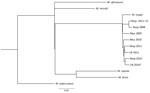
Figure. Maximum-likelihood single-nucleotide polymorphism (SNP) tree of 8 Mycobacterium orygis and 1 M. caprae isolates obtained from patients in New York, USA. Alignment of 5,242 total SNP positions was calculated by using...
M. orygis infection may be underreported in the literature because cases may be identified as MTB complex or misidentified as M. africanum or M. bovis (8). Through whole-genome sequencing, the New York State Department of Health identified 8 additional cases of M. orygis of 6,322 MTB complex isolates from New York tested (3 pulmonary, 2 lymph node, and 2 abscess samples) that were received during 2005–2016 but were initially misidentified (data not shown). All patients were from India, Pakistan, or Nepal and had moved to the United States. SNP analysis indicated that the M. orygis isolates were genetically similar, but all were distant from other members of the MTB complex (Figure) and contained the G1113A mutation in gyrB. The number of SNPs separating the 8 M. orygis isolates was 106–323, which excludes their belonging to an epidemiologic transmission cluster (9) and strongly suggests that the infections were independently acquired.
We found no previous reports of M. orygis originating in the Americas; the most notable epidemiologic risk factor in this patient was prior residency in India, where M. orygis was found in a variety of animals (10). Because all organisms in the MTB complex have a distinct host preference, it is possible that M. orygis is mostly present in animals and few cases occur in humans, similar to M. bovis. This case demonstrates the value of molecular methodologies such as whole-genome sequencing for providing more detailed insight into the clinical and epidemiologic aspects of the MTB complex.
Dr. Marcos is an associate professor of clinical medicine at Stony Brook University in New York, USA. His research interests include epidemiology of emerging infectious diseases, tickborne diseases, and global health.
References
- Brosch R, Gordon SV, Marmiesse M, Brodin P, Buchrieser C, Eiglmeier K, et al. A new evolutionary scenario for the Mycobacterium tuberculosiscomplex. Proc Natl Acad Sci U S A. 2002;99:3684–9. DOIPubMed
- van Ingen J, Rahim Z, Mulder A, Boeree MJ, Simeone R, Brosch R, et al. Characterization of Mycobacterium orygis as M. tuberculosis complex subspecies. Emerg Infect Dis. 2012;18:653–5. DOIPubMed
- Huard RC, Fabre M, de Haas P, Lazzarini LCO, van Soolingen D, Cousins D, et al. Novel genetic polymorphisms that further delineate the phylogeny of the Mycobacterium tuberculosis complex. J Bacteriol. 2006;188:4271–87. DOIPubMed
- Halse TA, Escuyer VE, Musser KA. Evaluation of a single-tube multiplex real-time PCR for differentiation of members of the Mycobacterium tuberculosis complex in clinical specimens. J Clin Microbiol. 2011;49:2562–7. DOIPubMed
- Shea J, Halse TA, Lapierre P, Shudt M, Kohlerschmidt D, Van Roey P, et al. Comprehensive whole-genome sequencing and reporting of drug resistance profiles on clinical cases of Mycobacterium tuberculosis in New York State. J Clin Microbiol. 2017;55:1871–82. DOIPubMed
- Lavender CJ, Globan M, Kelly H, Brown LK, Sievers A, Fyfe JA, et al. Epidemiology and control of tuberculosis in Victoria, a low-burden state in south-eastern Australia, 2005-2010. Int J Tuberc Lung Dis. 2013;17:752–8. DOIPubMed
- Dawson KL, Bell A, Kawakami RP, Coley K, Yates G, Collins DM. Transmission of Mycobacterium orygis (M. tuberculosis complex species) from a tuberculosis patient to a dairy cow in New Zealand. J Clin Microbiol. 2012;50:3136–8. DOIPubMed
- Tortoli E. Microbiological features and clinical relevance of new species of the genus Mycobacterium. Clin Microbiol Rev. 2014;27:727–52. DOIPubMed
- Walker TM, Ip CL, Harrell RH, Evans JT, Kapatai G, Dedicoat MJ, et al. Whole-genome sequencing to delineate Mycobacterium tuberculosisoutbreaks: a retrospective observational study. Lancet Infect Dis. 2013;13:137–46. DOIPubMed
- Thapa J, Paudel S, Sadaula A, Shah Y, Maharjan B, Kaufman GE, et al. Mycobacterium orygis-associated tuberculosis in free-ranging rhinoceros, Nepal, 2015. Emerg Infect Dis. 2016;22:570–2. DOIPubMed


.png)


No hay comentarios:
Publicar un comentario