Snapshots of Life: Color Coding the Hippocampus
The final frontier? Trekkies would probably say it’s space, but mapping the brain—the most complicated biological structure in the known universe—is turning out to be an amazing adventure in its own right. Not only are researchers getting better at charting the brain’s densely packed and varied cellular topography, they are starting to identify the molecules that neurons use to connect into the distinct information-processing circuits that allow all walks of life to think and experience the world.
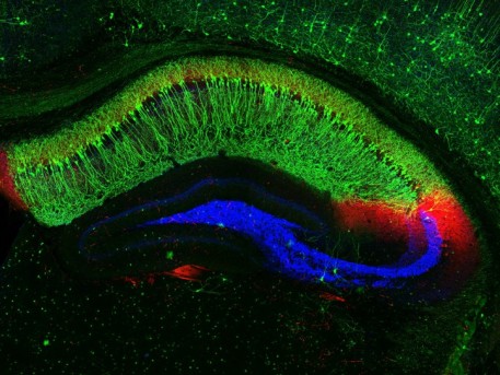
This image shows distinct neural connections in a cross section of a mouse’s hippocampus, a region of the brain involved in the memory of facts and events. The large, crescent-shaped area in green is hippocampal zone CA1. Its highly specialized neurons, called place cells, serve as the brain’s GPS system to track location. It appears green because these neurons express cadherin-10. This protein serves as a kind of molecular glue that likely imparts specific functional properties to this region. [1]
In red is hippocampal zone CA2. It’s important for forming memories of social interactions. These nerve cells harbor a very different set of proteins that give them distinct properties from the place cells. In fact, the red staining indicates the presence of a protein, RGS14, that’s uniquely made by CA2 neurons.
The swoosh of blue shows the transmission sites of nerve signals between neurons in the neighboring CA3 zone and dentate gyrus, part of the hippocampus involved in episodic memories. This signaling is thought to be necessary for pattern separation, which is the ability to distinguish between closely related memories. At the top right, you can see a portion of another brain region, the cerebral cortex. Some of the neurons there also appear green because they too express cadherin-10.
This image comes from Raunak Basu, a recently graduated Ph.D. student in the NIH-supported lab of Megan Williams at the University of Utah School of Medicine, Salt Lake City. As Basu notes, less is always more when photographing any of the cell-dense regions of the brain that block and badly distort light. So Basu used this 100-micron (0.0039 inches) thick slice of a mouse brain, which thinned the volume of cells, and placed it under a confocal microscope.
Pleased with the striking color and details, Basu snapped this photo and named it “Rainbows in Our Mind,” a reference to the color-coded cell types in the image and their contribution to thought and memory. It was one of the winners in the University of Utah’s 2016 Research as Art competition.
The image also drives home the point that neuronal connectivity is exquisitely precise, which is highlighted by the blue nerve terminals stopping precisely before entering the green zone. With tens of trillions of neuronal connections in the human brain, understanding the molecules that drive the connectivity process will go far in the quest to explore this profoundly complex next frontier.
Reference:
[1] The classic cadherins in synaptic specificity. Basu R, Taylor MR, Williams ME. Cell Adh Migr. 2015;9(3):193-201.
Links:
Williams Lab (University of Utah, Salt Lake City)
Research as Art (University of Utah)
NIH Support: National Institute of Mental Health


No hay comentarios:
Publicar un comentario